
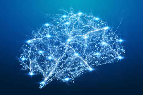
Research techniques in the field of neuroscience are extremely important. This is especially true when you consider what’s expected in the coming years for this particular science. As a matter of fact, it was only a short time ago that neuroimaging techniques were only a dream. This meant that certain theories about the nervous system were impossible to establish.
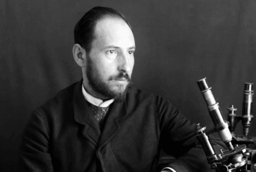
In 1988, the Spanish physician Santiago Ramon y Cajal described the morphology of neurons through a complex system of staining. Since then, there have been several milestones in neurobiology. For example, the discovery of the electroencephalogram (EEG) and the classification of the biological rhythms of the brain.
Research tools in neuroscience
Neuroscience research tools are devices that allow scientists to study the nervous system. So-called neuroimaging techniques are used for both clinical and academic purposes. These tools allow the nervous system to be approached in four different ways:
- Functional. Describes the function of the nervous system. It does this through flow rate or the degree of activation of various zones.
- Structural. Delivers anatomical information about the brain or other structures of the nervous system.
- Electrical. Delivers information about the electrical activity of the nervous system.
- Stimulation. Non-invasive stimulation of the brain.
Techniques of structural neuroimaging in neuroscience research
Among the structural techniques specialized in providing anatomical information, the most basic and widely used is radiography. This technique consists of the emission of X-rays. Depending on the density of the tissue, a different image is projected onto the photographic plate. There are two types of X-rays:
- Common radiography. This is only used for X-rays. In addition, it’s commonly used to obtain information about the bones that surround the nervous system. For example, in the case of fractures.
- Contrast radiography. It’s also used with X-rays, but it involves inserting a catheter through the femoral artery. The hyperdense contrast allows the detection of vascular alteration.
Computerized axial tomography
This is the well-recognized scanner in the shape of a tube. It uses the emission of X-rays from different directions, thus providing a more complete image. It’s a rapid technique with a low cost that detects tumors, aneurisms, and hemorrhages. On the other hand, radiation isn’t good for the body. Furthermore, the definition isn’t as high as the other techniques.
Magnetic resonance
This research technique in neuroscience picks up images of the interior of the body with elevated resolution and in a safe way. However, it needs to be conducted with care as it’s incompatible with any kinds of metal implants in the body. It’s based on the phenomena of nuclear magnetic resonance. In fact, the machine registers the signals of radiofrequency emitted through the hydrogen atoms, previously subjected to a magnetic field.
Hydrogen atoms are abundantly present in the body. Because of this, magnetic resonance is a high-resolution technique. What’s more, there are no harmful effects and it doesn’t use radiation. On the other hand, it’s extremely expensive.
Tractography
This is a tool that uses magnetic resonance to evaluate the areas of white matter. Actually, these mainly consist of water and are in charge of delivering nerve information at high speed. They’re called myelinated axons. The technique is able to evaluate the subcortical structure of the brain, allowing the detection of neurodegenerative diseases and epilepsy.
Techniques of functional neuroimaging in neuroscience research
These are the tools of studies in neurosciences. They detect the changes in live brain activity. Experts tend to use these methods to evaluate cognitive processes in combination with the functioning of their anatomical correlates.
Positron emission tomography (PET)
This technique is based on the introduction of radioactive substances into the bloodstream. Cells with high metabolic activity absorb the substances. In this process, the isotopes that emit positrons cancel out when they combine with electrons, generating electromagnetic energy, which is captured by the device.
PET is a widely used technique to detect brain tumors since they tend to have a higher metabolism. In turn, it’s also used to detect neurodegenerative diseases. For example, in the case of Alzheimer’s, the degeneration of cells won’t cause as much substance to be absorbed. Hence, the image will differ from that of a normal brain.
Single-photon emission computed tomography
This is a recording technique that’s similar to the previous one. However, it uses gamma radiation, produced by an isotope directly from within the body. Furthermore, it requires a receptor, through which an image will be generated. It highlights in colors the different degrees of brain activation.
Functional magnetic resonance
This is an MRI where, at the same time, some cognitive activity is carried out. It’s based on the fact that the neurons involved in a mental process will need more energy and therefore more oxygen from the blood. In fact, when we perform mental tasks, our consumption of oxygenated blood (which has magnetic properties) increases. That activity is recorded by the device. Despite having a high cost, it allows the location of cognitive functions in the brain.
Electrophysiological techniques
These techniques allow for registering the electrical activity of the brain. For example:
- Electroencephalogram. It measures the electrical power of the brain. In addition, it also measures the type of wave and frequency in which it’s functioning.
- Electromyogram. It evaluates the electric activity of the muscles. It’s used for the exploration of the peripheral nerves.
- Electrooculogram. It measures the electrical muscle recording of the eyes and the sleep phases.

Techniques of cerebral stimulation
These techniques allow brain activity to be influenced by two types of stimuli:
- Magnetic. With transcranial magnetic stimulation, a current is safely induced in the brain. This induction is achieved by a flow of current that passes through a coil and generates a magnetic field.
- Electrical: This uses a low-intensity current by means of electrodes on the scalp. As a result, the induction causes changes in the excitability of the neurons of the cortex.
These forms of stimulation tend to only reach the areas of the cortex. In fact, they’re mostly used to identify mental processes, as well as to create virtual injuries. Their application to improve performance or treat disorders such as phobias is still under investigation. Furthermore, they shouldn’t be used for people with epilepsy, implants, or pregnant women.
The importance of neuroscience research techniques
The diseases that affect the nervous system can have serious consequences. Therefore, the timely detection of a tumor or hemorrhage is of utmost importance in increasing the patient’s chances of survival. At the same time, detecting a neurodegenerative disease in its first phases is key in delaying its symptoms.
Furthermore, scientific progress has allowed scientists to delve into brain function. For example, it’s currently possible for them to compare the brain of a depressed person with that of a normal person, obtaining functional differences associated with symptoms. In the same way, they can identify brain areas and processes corresponding to a particular function, such as attention.
The post Neuroscience Research Techniques appeared first on Exploring your mind.


Comments